Diverticulosis vs. Diverticulitis: Debunking the Seeds & Nuts Myth, Dietary Protocols, & Colonoscopy Timing
A gastroenterological evaluation of colonic outpouchings. Why historical prohibitions against nuts, seeds, and popcorn are medically unfounded, how modern guidelines manage uncomplicated flares without routine antibiotics, and when to safely perform surveillance colonoscopy.
Key Clinical Practice Takeaways
- Anatomical Distinction: Diverticulosis is the quiescent presence of structural pseudodiverticula herniating through muscular gaps at penetrating vasa recta sites. Diverticulitis is an acute micro-perforation event triggering focal necrosis and pericolic inflammation.
- Myth Completely Debunked: The prospective 18-year Harvard Health Professionals study (Strate et al., JAMA 2008; PMID: 18728264) showed nuts and popcorn intake is associated with a 20–28% lower risk of diverticulitis, conclusively overthrowing decades of seed avoidance.
- Selective Antibiotic Stewardship: Current American Gastroenterological Association (AGA) and European WSES guidelines endorse supportive outpatient care without routine antibiotics for mild, uncomplicated diverticulitis in immunocompetent patients.
- Surveillance Colonoscopy Window: Colonoscopy is strictly contraindicated during the acute flare due to perforation risk, but must be scheduled 6 to 8 weeks post-resolution to definitively exclude occult colonic adenocarcinoma mimicking inflammatory phlegmon.
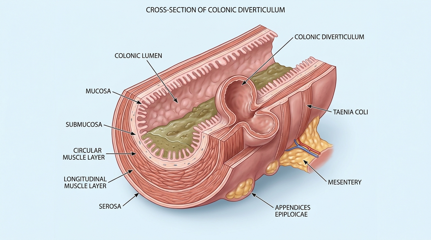
1. Structural Anatomy: Pulsion Herniation and the Vasa Recta
Diverticular disease represents one of the most prevalent anatomical findings encountered during routine screening colonoscopy in Western nations, affecting over 50% of adults over age 60 and more than 65% of individuals past age 80 (Peery et al., Gastroenterology 2022; PMID: 34678258).
To understand this condition, one must distinguish between true and false diverticula. A true diverticulum (such as a congenital Meckel's diverticulum) involves herniation of all four wall layers: mucosa, submucosa, muscularis propria, and serosa. In contrast, colonic diverticula are "pseudodiverticula" (false diverticula).
The human colon wall features three longitudinal muscle ribbons called taeniae coli overlying a continuous circular muscular coat. To supply oxygen and nutrients to the rapidly dividing colonic mucosal lining, mesenteric arterial branches known as vasa recta must pierce through this circular muscle layer. These vascular penetration points create microscopic focal gaps in the muscular defense.
Over decades of low-fiber dietary intake, reduced stool bulk forces the colon to generate powerful segmental peristaltic contractions to propel feces forward. According to Laplace's Law (Wall Tension = Pressure × Radius), the narrow lumen of the sigmoid colon experiences the highest intracolonic pressures in the digestive tract. These high hydraulic pressures push the inner mucosa and submucosa outward through the vasa recta gaps like an inner tube bulging through a weak tire wall. This pulsion process leaves the resulting diverticular pocket covered only by delicate mucosa and outer serosa, directly abutting mesenteric fat.
2. Diverticulosis vs. Diverticulitis: The Clinical Chasm
Patients frequently conflate these two conditions, yet their clinical implications are entirely distinct:
| Diagnostic Parameter | Diverticulosis | Diverticulitis |
|---|---|---|
| Pathology Definition | Structural presence of non-inflamed pseudodiverticula | Acute inflammation, focal necrosis & micro-perforation |
| Typical Symptoms | 100% Asymptomatic in ~80–85% of individuals | Constant left lower quadrant (LLQ) pain, fever, leukocytosis, nausea |
| Primary Etiology | Age-related collagen elastosis + high intracolonic pressure | Inspissated fecalith impaction → ischemia → bacterial translocation |
| Diagnostic Standard | Routine optical colonoscopy or incidental CT | Abdominopelvic CT with IV contrast (fat stranding, wall thickening) |
| Lifetime Progression Risk | Only 4% to 7% ever progress to acute diverticulitis | Recurrence risk is ~20% at 5 years and ~35% at 10 years |
Diverticulitis is not simple luminal inflammation; it is an extraluminal process. When an inspissated fecalith becomes lodged inside the narrow neck of a diverticulum, mucosal abrasion impairs venous return, leading to localized ischemic necrosis. The ultrathin fundus of the diverticulum suffers a micro-perforation, allowing enteric bacteria to leak into surrounding pericolic fat and peritoneal connective tissue.
3. The Debunked Dietary Myth: Seeds, Nuts, and Popcorn
For over half a century, millions of individuals diagnosed with diverticulosis were given rigid dietary restrictions: "Never consume nuts, popcorn, sesame seeds, poppy seeds, strawberries, cucumbers, or tomatoes. The sharp fragments will fall into your diverticula and cause an acute rupture."
This dogma was based entirely on plausible-sounding anecdotal speculation without a shred of empirical scientific evidence. In 2008, the medical community witnessed the definitive collapse of this myth.
In a landmark prospective cohort investigation published in The Journal of the American Medical Association (JAMA), Dr. Lisa L. Strate and researchers from Harvard Medical School followed 47,228 men from the Health Professionals Follow-Up Study over an 18-year period (Strate et al., JAMA 2008; PMID: 18728264). Men who regularly ate nuts, seeds, and popcorn were tracked for incident diverticulitis and diverticular bleeding.
The results stunned traditional clinicians:
- No Increased Risk: There was zero statistical association between the consumption of nuts, corn, seeds, or popcorn and the development of diverticulitis or diverticular hemorrhage.
- Statistically Significant Protection: Men with the highest nut consumption (≥ 2 times per week) experienced a 20% reduction in diverticulitis risk (Hazard Ratio 0.80; 95% CI 0.63–1.01) compared to men who avoided nuts.
- Popcorn Protective Effect: Men consuming popcorn ≥ 2 times per week had a 28% reduced risk of diverticulitis (Hazard Ratio 0.72; 95% CI 0.56–0.92) compared to those consuming it less than once monthly.
Why would nuts and popcorn reduce diverticulitis? Because whole nuts, seeds, and popcorn hulls are rich in soluble and insoluble dietary fiber, plant polyphenols, and micronutrients that improve stool consistency, optimize microbial diversity, and lower intracolonic intraluminal pressure.
Subsequent formal clinical practice guidelines from the American Gastroenterological Association (Stollman et al., Gastroenterology 2015; PMID: 26453777) explicitly state: "The AGA suggests against advising patients with a history of diverticulitis to avoid the consumption of seeds, nuts, berries, or popcorn." Telling patients to eliminate these healthy dietary staples is an outdated relic of obsolete medical training.
4. Evidence-Based Nutritional Management Across Disease Phases
Nutritional recommendations must be strictly tailored depending on whether a patient is in a quiescent state of diverticulosis or recovering from an acute inflammatory flare:
Phase 1: Quiescent Diverticulosis (Long-Term Prevention)
The primary objective is maximizing stool bulk to prevent segmentation and lower intracolonic pressure. Target 25 to 35 grams of dietary fiber daily, combining both soluble and insoluble sources:
- Soluble Fiber (Mucilaginous Gel-Forming): Oats, psyllium husk, ground flaxseeds, chia seeds, lentils, apples. Softens stool and binds water.
- Insoluble Fiber (Structural Bulk): Wheat bran, dark cruciferous vegetables, nuts, root vegetables. Accelerates colonic transit and eliminates hard, dry fecaliths.
- Adequate Hydration: 2 to 3 liters of water daily to support fiber expansion.
Phase 2: Acute Diverticulitis Flare (Resting the Bowel)
During an acute inflammatory attack, high-fiber foods must be temporarily withheld because active peristalsis and rough bulk can aggravate the inflamed, micro-perforated segment:
- Days 1–3: Clear liquid diet (broth, electrolyte solutions, water, clear herbal teas) to minimize luminal pressure and stool volume.
- Days 4–10: Transition to a temporary low-residue, low-fiber diet (plain white rice, poultry, white toast, eggs, canned peeled peaches).
- Post-Flare Recovery: Gradually re-titrate dietary fiber over 4 to 6 weeks back to 30 grams per day as mucosal inflammation completely resolves.
5. Modern Medical Therapy: Selective Antibiotic Stewardship
Historically, every patient presenting with CT-proven diverticulitis was automatically prescribed broad-spectrum antibiotics (typically ciprofloxacin and metronidazole, or amoxicillin-clavulanate).
Over the past decade, large European randomized clinical trials (the AVOD trial and DIABOLO trial) evaluated observational supportive care vs. antibiotic therapy in mild, uncomplicated diverticulitis (defined as localized pericolic fat stranding without abscess, extraluminal free air, fistula, or obstruction). These trials demonstrated no significant difference in time to recovery, rate of complications, or recurrent attacks between patients treated with antibiotics and those treated with supportive analgesia alone.
Current AGA guidelines recommend that antibiotics be utilized selectively rather than routinely in immunocompetent outpatients presenting with mild, uncomplicated diverticulitis. Antibiotic therapy is reserved for patients exhibiting systemic toxicity (temperature > 38.5°C, marked leukocytosis > 15,000/μL), immunosuppression, severe comorbidities, or CT evidence of complicated disease (abscess ≥ 3 cm, macro-perforation).
6. Colonoscopy Timing: The Critical 6-to-8-Week Interval
A vital principle in gastroenterology is the timing of endoscopic visualization following diverticulitis:
Endoscopic evaluation is strictly contraindicated during an acute flare. Insufflating carbon dioxide or air into a lumen with an actively micro-perforated, ischemic, edematous diverticulum can blow open a contained perforation into catastrophic free peritonitis requiring emergent colostomy surgery.
However, a diagnostic colonoscopy must be performed 6 to 8 weeks after complete clinical resolution of the acute episode (if the patient has not undergone a high-quality screening colonoscopy within the preceding 12 to 24 months).
Why is this follow-up mandatory? Because an infiltrating colorectal adenocarcinoma can closely mimic acute diverticulitis on cross-sectional CT imaging. Both conditions present with focal colonic wall thickening, pericolic soft-tissue stranding, and luminal narrowing. Multiple clinical series demonstrate that between 1.5% and 5% of patients diagnosed with acute diverticulitis—and up to 11% of patients with complicated diverticulitis—are found to harbor an underlying malignant tumor upon follow-up optical colonoscopy. Waiting 6 to 8 weeks permits full resolution of benign inflammatory swelling, allowing the endoscopist to safely navigate the sigmoid colon and visualize the mucosal architecture.
7. Peer-Reviewed Clinical Citations (PubMed References)
1. Strate LL, Liu YL, Syngal S, et al. Nut, corn, and popcorn consumption and the incidence of diverticular disease. JAMA. 2008;300(8):907-914. PMID: 18728264
2. Stollman N, Smalley W, Hirano I, et al. American Gastroenterological Association Institute Guideline on the Management of Acute Diverticulitis. Gastroenterology. 2015;149(7):1944-1949. PMID: 26453777
3. Peery AF, Crockett SD, Murphy CC, et al. Burden and Cost of Gastrointestinal, Liver, and Bile Diseases in the United States: Update 2021. Gastroenterology. 2022;162(2):621-644. PMID: 34678258
4. Sartelli M, Weber DG, Kluger Y, et al. 2020 update of the WSES guidelines for the management of acute colonic diverticulitis in the emergency setting. World J Emerg Surg. 2020;15(1):32. PMID: 32381121
Frequently Asked Questions: Diverticular Health
Can eating nuts, seeds, strawberries, or popcorn trigger diverticulitis?
No. The long-standing medical myth that sharp seeds or nut fragments lodge in diverticular pockets has been completely disproven. A landmark 18-year prospective study published in JAMA by Dr. Lisa Strate and colleagues (PMID: 18728264) following 47,228 men demonstrated no association between nut, corn, or popcorn consumption and diverticulitis. In fact, frequent nut and popcorn consumers exhibited a 20% to 28% reduced risk of diverticular inflammation compared to non-consumers. Professional guidelines from the American Gastroenterological Association (AGA) explicitly advise against restricting seeds and nuts.
What is the physical difference between diverticulosis and diverticulitis?
Diverticulosis is the structural presence of harmless, non-inflamed outpouchings (pseudodiverticula) where colonic mucosa herniates through weaknesses in the circular muscularis propria. It is asymptomatic in over 80% of individuals. Diverticulitis occurs when an outpouching suffers micro-perforation, focal tissue ischemia, bacterial translocation, and acute pericolic inflammation, producing severe abdominal pain, fever, leukocytosis, and localized tenderness.
Why is a colonoscopy scheduled 6 to 8 weeks after an acute flare of diverticulitis?
Performing a colonoscopy during an active episode of acute diverticulitis is contraindicated due to high risk of bowel perforation from mechanical endoscope manipulation and air insufflation into inflamed tissue. However, scheduling an optical colonoscopy 6 to 8 weeks after symptoms completely resolve is crucial to rule out occult colorectal cancer or advanced adenomatous polyps, which can mimic diverticular inflammation or luminal strictures on emergency abdominal CT imaging.